Thigh muscles are responsible for allowing normal gait and proper lower extremity function 1. He caring foot doctors and staff at AllCare Foot Ankle Female Podiatrist believe in making quality podiatry services available to all patients including those without insurance.
Radiologist Foot X Ray Anatomy Facebook

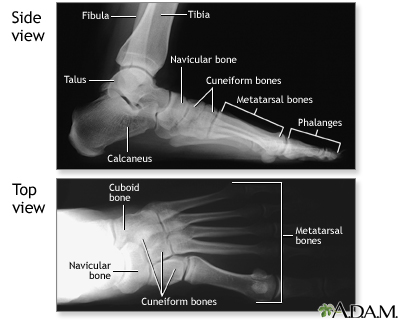
Normal Foot X Ray Medlineplus Medical Encyclopedia Image
Epos Trade
Most land mammals including humans have a 2-3-3-3-3.

Foot x ray anatomy. Ii Midfoot navicular cuboid and cuneiforms. This digital content included in this resource is licensed under the Creative Commons Attribution-NonCommercial-NoDerivatives 40 International CC BY-NC-ND 40 It is the users responsibility to read understand and abide by the terms and conditions under which they can access and use the licensed content. Non-contrast coronal CT head.
In this article we shall look at the anatomy of the bones of the foot their bony landmarks articulations and clinical correlations. Coxa vara is a deformity of the hip whereby the angle between the head and the shaft of the femur is reduced to less than 120 degrees. This article lists a series of labeled imaging anatomy cases by system and modality.
The podiatrists at Reston Manassas and Leesburg Foot and Ankle Centers provide full adult and pediatric foot and ankle services including treatment for heel pain plantar fasciitis bunions warts fungal nails ingrown nails fractures sprains sports injuriesneuromasas well as diabetic foot care custom orthotics Medicare approved diabetic shoes foot surgery and ankle surgery. Other tests your doctor may run include. Angiogram axial CT head.
A doctor may look for swelling deformity pain discoloration or skin changes to help diagnose a foot problem. The radiation is created when an electric current is generated from a high voltage generator causing electrons to boil-off from the cathode end of an X-ray tube assembly. In 1858 Drs Henry Gray and Henry Vandyke Carter created a book for their surgical colleagues that established an enduring standard among anatomical texts.
The foot can also be divided up into three regions. Pseudofracture of the apophysis is a partially ossified apophyseal plate that appears on an X-ray as if it was a fracture. If more detail is needed however an orthopedic doctor will likely want to do magnetic resonance imaging.
And iii Forefoot metatarsals and phalanges. A plain X-ray film may identify problems with the bones or ankle joint but it cannot diagnose Achilles tendon problems. Building on over 160 years of anatomical excellence In 1858 Drs Henry Gray and Henry Vandyke Carter created a book for their surgical colleagues that established an enduring standard among anatomical texts.
Blood tests which can identify conditions such as gout. The central ray is a simplified way of indicating the direction in which an x-ray beam travels. This ankle and foot examination OSCE guide provides a clear step-by-step approach to examining the ankle and.
The distal phalanges of ungulates carry and shape nails and claws and these in primates are referred to as the ungual phalanges. Susan Standring MBE PhD DSc FKC Hon FAS Hon FRCS Trust Grays. Building on over 160 years of anatomical excellence.
Non-contrast sagittal CT head. This results in the leg being shortened and the development of a limpIt may be congenital and is commonly caused by injury such as a fracture. History of phalanges Etymology.
Angiogram coronal CT head. Elizabeth Quinn is an exercise physiologist. Thigh Magnetic Resonance Imaging.
After more than 160 years of continuous publication Grays Anatomy remains the definitive comprehensive reference. This is a normal variant of the calcaneus and does not require treatment. Susan Standring MBE PhD DSc FKC Hon FAS Hon FRCS.
Loss of joint alignment can represent. Anterior-posterior denotes that the central ray passes first through the anterior anatomy and exits posteriorly while the posterior-anterior denotes that the central beam passes from posterior to anterior. Clinical Examination A comprehensive collection of clinical examination OSCE guides that include step-by-step images of key steps video demonstrations and PDF mark schemes.
The term phalanx or phalanges refers to an ancient Greek army formation in which soldiers stand side by side several rows deep like an arrangement of fingers or toes. We understand many patients have questions about the cost of a consultation and services without insurance and our friendly billing support team is here to answer your questions. A standard X-ray can confirm a bone fracture or arthritis damage.
A plain X-ray film of the feet can detect. Radiology Department of the Rijnland Hospital Leiderdorp the Netherlands. The Anatomy of the Foot and Common Foot Problems By.
If your doctor suspects a broken bone fracture or bone spurs theyll order an X-ray of the foot. On a chest x-ray lung abnormalities will either present as areas of increased density or as areas of decreased density. When checking any post-traumatic foot X-ray it is crucial to assess alignment of the bones at the joints.
Continued Achilles Tendon Treatments. Different projections describe how the central ray travels through anatomy. Upper ankle joint tibiotarsal talocalcaneonavicular and subtalar jointsThe last two together are called the lower ankle joint.
A collection of anatomy notes covering the key anatomy concepts that medical students need to learn. The medial thigh muscles are responsible for the adduction movement of a. Ankle and foot examination frequently appears in OSCEs and youll be expected to identify relevant clinical signs using your examination skills.
Plain film x-ray is the most common diagnostic radiological modality used in hospitals today. X-ray of the chest also known as a chest radiograph is a commonly used imaging study and is the most frequently performed imaging study in the United StatesIt is almost always the first imaging study ordered to evaluate for pathologies of the thorax although further diagnostic imaging laboratory tests and additional physical examinations may be necessary to help confirm the diagnosis. I Hindfoot talus and calcaneus.
Congenital tarsal coalition is a connection between tarsals usually the calcaneus and talus that prevent the tarsals from articulating properly. The ankle joint also known as the talocrural joint allows dorsiflexion and plantar flexion of the footIt is made up of three joints. Non-contrast axial CT head.
The thigh has some of the bodys largest muscles.

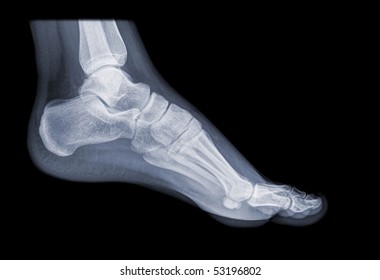
X Ray Foot Images Stock Photos Vectors Shutterstock

Foot Radiograph Anatomy Quiz Radiology Case Radiopaedia Org

Radiographic Anatomy Of The Foot Youtube

Medial And Top View X Ray Of Bones The Of Foot With Sesamoid Stock Vector Illustration Of Health Drawing 162518607

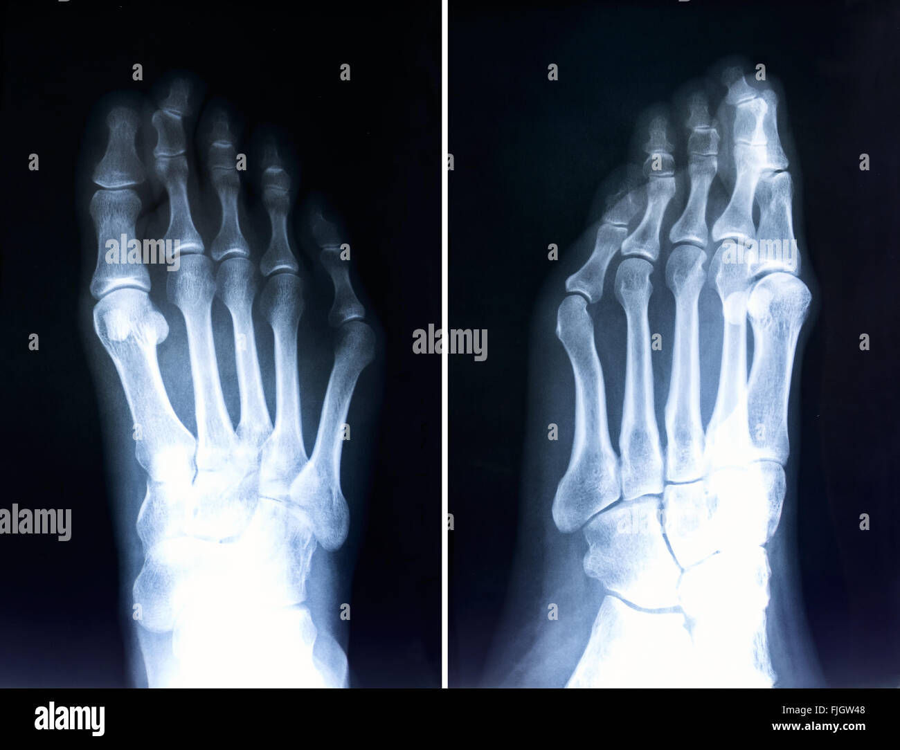
Human Foot X Ray High Resolution Stock Photography And Images Alamy

Radiograph Of A 3 Year Old Child S Right Foot The Bmj

Anatomy Of The Bones Of The Foot The Bmj

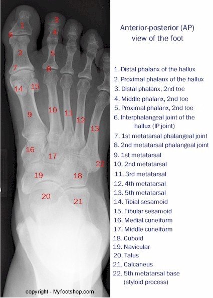
X Ray Of The Foot Anterior Posterior View Myfootshop Com


